This is a plain-language summary of the article “A Simple Preparation Method for Full-Range Electron Tomography of Nanoparticles and Fine Powders” in the journal Microscopy and Microanalysis
To really understand how something works, you want to be able to see it from all directions, and even look inside it. A great way to do this is by taking a 3D image.
Doctors do this using a “CT scan” when they need to look inside a patient (without cutting them open) and figure out what might be going wrong in the complicated innards of the human body. The CT (computed tomography) scan works by taking X-ray images of the patient from all directions, spinning an X-ray source and a camera around them in a circle. A computer then uses those images to work backward and calculate a 3D image of the inside of the patient.
Nanoscientists work with objects and materials that are extremely small – smaller than the cells in your body, and sometimes only a few atoms across – which behave in unique ways because of their small size. To understand these nano-things and use them to build useful structures and devices, nanoscientists also sometimes need to see them in 3D.
Seeing things this small is quite a bit harder than seeing things the size of a person. Nano-things are too small even to see with an ordinary microscope (the kind you might have used in a biology class) since they are smaller than the wavelength of visible light. This means light waves will lose all of the details when they bounce off and form an image.
Instead, scientists often use electron microscopes, which replace the visible light in an ordinary microscope with a beam of fast-moving electrons. These microscopes are great for imaging nano-things, and powerful ones can even resolve individual atoms.
But for complicated nano-objects, a single flat picture is often not enough. Fortunately, an electron microscope can also be used to take 3D images of nano-things like the CT scans of the body used in medicine. This is called “electron tomography”, and works in a similar way. The electron microscope takes images of the object as the object spins around an axis, and the images are used to put together the 3D image.
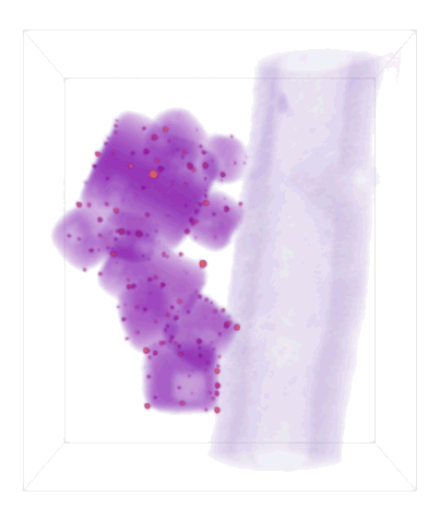
Doing tomography in an electron microscope has some drawbacks compared to CT using X-rays. The object of interest has to be held and rotated somehow. Typically it sits on a thin sheet of carbon stretched over a disk-shaped metal grid, which is held at the end of a metal rod called a holder. But electrons can’t travel through very much stuff without getting blocked. So when a researcher tries to spin an object around in the electron microscope to do tomography, some views are blocked by the grid or the sides of the holder. So the researcher doesn’t get to see the object from every angle, and this means that some of the information about it is missing. This degrades the quality of the 3D image by making it blurred in one direction and distorting its shape. These problems can make it hard to interpret the 3D image or make accurate measurements.
In recent years, researchers have gotten around this problem by making their material of interest into a needle shape so that it can rotate all the way around without getting blocked. This gives great quality 3D images, but to make a needle for tomography you have to carved it out and sharpen it with a beam of fast-moving particles (kind of like cutting with a water jet). This works for some materials, but it’s difficult and doesn’t work well if you want to look at a whole object without cutting it.
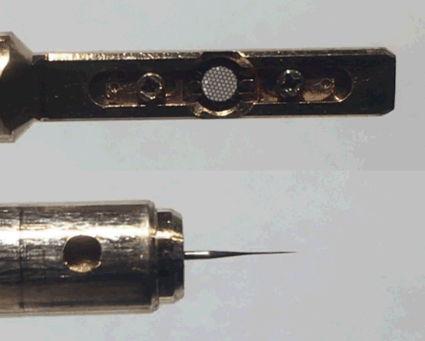
My co-workers and I developed a new approach: instead of carving out sample, we start with a thin carbon nano-fiber on the end of a needle and collect our nano-objects on the fiber. The fibers are thin and low-density, so electrons can easily pass through to give a clear view of the objects of interest when they are rotated all the way around.
This approach (“particles on a stick” to a state-fair-goer) helps make clearer, more accurate 3D images to help nanoscientists make sense of their tiny worlds.